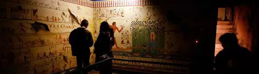
Latest News: Full Story
|
||||||||||||||||||
|
Today in Silicon Valley, a team of world-renowned experts proved those parents right although the mummy’s high-tech resurrection may not quite be what ancient Egyptians had in mind.
Recently, researchers allowed the public to literally come face to face with the rare mummified remains of the ancient Egyptian child. Equipped with the most detailed 3D models ever created of a mummy, a team of experts showed how 60,000 exceptionally high-resolution 2D scans helped give life to the mummy without disturbing its delicate form.
The result is the highest quality interactive visualization of a mummy ever seen one that allowed specialists in various fields from Stanford University School of Medicine and the Stanford-NASA National Biocomputation Center to arrive at several conclusions about the child who lived and died 2,000 years ago.
Curators at the Rosicrucian Egyptian Museum & Planetarium, which has housed the mummy since about 1930, have named the child Sherit, an ancient Egyptian name that means “little one.”
History-making scan and visualization
For the project, radiologists at Stanford University School of Medicine used an AXIOM Siemens scanner, one of only five CT scanners in the world capable of producing such high-resolution images. Stanford Radiology’s state-of-the-art scanner generated 2D slices as thin as 200 microns several times thinner than the 750-micron slices used to create the popular 3D visualization of King Tutankhamen’s mummy. In fact, at 92 gigabytes, Stanford Radiology’s child mummy scans generated nearly 35 times more information than the scans conducted on King Tut.
To combine that information into a fully interactive 3D model of the entire mummy and its contents, researchers relied on the powerful Silicon Graphics computers running specialized software from Germany’s Volume Graphics GmbH. With the software's real-time ray tracing technology similar to that used to create hit animated motion pictures researchers were able to generate a 3D model of incomparable quality and fidelity.
After conducting detailed analyses of several areas including the hands, teeth, feet, skull, groin, spine and chest plate researchers were able to arrive several conclusions about the mummy. Among them:
“Real anatomy exists in three dimensions, so any time you can view anatomical data in 3D, you’ll have a much more accurate picture of the subject,” said Paul Brown, DDS, of the Stanford-NASA National Biocomputation Center. Brown and a team of fellow dentists, orthodontists and oral surgeons determined the mummy’s age and other features by studying the 3D visualization. “Even multiple two-dimensional CT slices can never allow you to understand a subject’s dental condition as quickly or as accurately as a quality 3D visualization.”
According to Brown, high-resolution scanning and visualization technology already is transforming medical, dental and orthodontic procedures, with specialists using them to speed diagnoses, plan surgeries and predict growth patterns. Technologies like the ones used on the child mummy will only accelerate those advances.
“I’ve worked with high-resolution 3D visualizations for years,” added Brown, who has performed more than 35,000 root canals and today conducts research at Stanford and teaches at two other California universities. “By far, this is the best visualization I’ve ever seen. There is no comparison.”
“Mummy visualizations are certainly growing more prevalent, but in terms of enabling technology, nothing else comes close to the quality, resolution and interactivity that we’ve achieved with SGI visualization systems,” said Lisa Schwappach-Shirriff, curator, Rosicrucian Egyptian Museum and Planetarium, which previously had relied on X-rays taken in the 1960s for information on the child mummy. “With SGI making historic use of higher-resolution scans and volume visualization applications, scientists were able to model Sherit with unprecedented realism. The images of this little girl are breathtaking, and the details that we can see on her are nothing short of amazing.”
Team brings child mummy to life
Visitors to the Rosicrucian Egyptian Museum are now able to peer across 2,000 years of history to see the girl’s face as real as life itself. After digitally modeling her skull from CT data, a team of scientists led by reconstructive surgeon Stephen Schendel, MD, DDS, professor of surgery at Stanford, displayed a physical replica precisely constructed to match the girl’s actual skull. Using that physical model along with clues derived from studying one of her still-intact ears and knowledge of facial characteristics common to Egyptian children, the team created a clay bust of the little girl’s face.
“The bust brings to life the story of this little girl who lived at a time when Egyptians, Romans, Jews and Christians all lived side by side,” said Schwappach-Shirriff. “This mummy is no longer just a fascinating artifact, but a lively young child who lived many ages ago.”
A film and interactive display of the incredible results of this scanning are on display until September 5 in the "Egypt through Time" exhibition. The display includes a forensic recreation of the child's face and head and an interactive exhibit that allows visitors to uncover the mummy for themselves.
The news about the results of this study of the Museum's child mummy was released last week at a media conference. At the end of the presentation, the press corps applauded, which one of the representatives from a local TV station said "almost never happens!" Since then, publicity has been building, on TV, Radio, print media and the Internet. Silicon Graphics estimates that over 190 million people have been exposed to the story through all media so far. The story is going out over the Associated Press, and is featured in Wired News Online and Time Magazine. View an abstract of the Time article at Time Magazine online.
Subscribe to our email newsletter